Child Safety Week highlights after-school road accidents – and much more!

As part of Child Safety Week (June 1-7), the Child Accident Prevention Trust (CAPT) has revealed findings that suggest the end of the day is when serious accidents and injury happen to children – especially on the road.

Teatime spells the start of danger for children, with over half of all serious road accidents occurring between 3pm and 7pm.

The peak in child road deaths and injuries is linked to the after-school rush. CAPT reports that there are more serious and fatal injuries to school-age pedestrians in the afternoon and early evening than at any other time of day.

Over half of police-reported child pedestrian fatal or serious road injuries occur between 3pm and 7pm among children and young people under 16. Injuries reach their peak from 3pm to 4pm. In the five years from 2008-2012, 3,500 children were killed or seriously injured on the roads between 3pm and 7pm, that is 13 children every week.

Katrina Phillips, Chief Executive for CAPT, says: “Parents are up against it to get everyone home, tea on the table, clothes ironed and tired children into the bath. It’s hardly surprising safety measures get missed.

“But these can be devastating injuries. A child can suffer brain damage if hit by a car. Simple changes to teatime routines can protect children from serious harm – [such as] practising road safety on the walk home from school.”

Dianne Yates, Partner and Head of Serious and Catastrophic Injury for Birchall Blackburn Law, says: “A split second lapse in safety can have a life changing effect, not just on the child’s future but on the whole family. We help injured children and their families in the aftermath of an accident and we see the consequences first hand. It can be utterly devastating. We cannot stress enough the importance of initiatives like Child Safety Week and the work of CAPT to prevent injury in the first place – hopefully avoiding the need for our later involvement.”

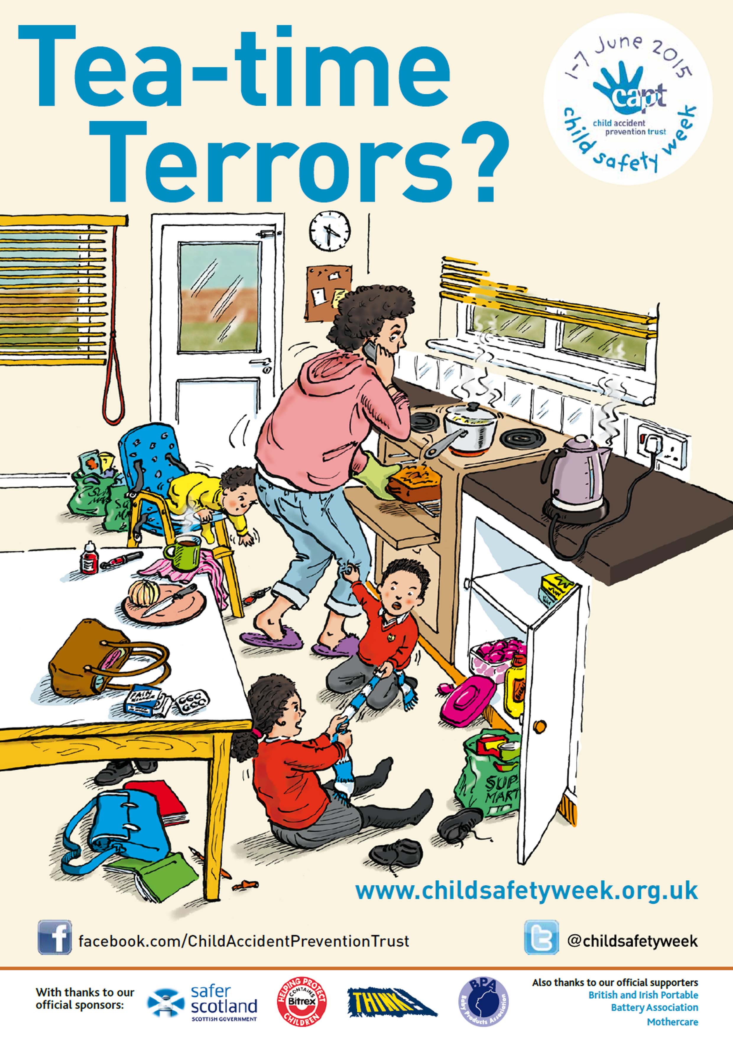
As part of Child Safety Week, CAPT is highlighting lots of potential accidents that can happen to children during the often stressful teatime period. As well as the dangers of the road, children are more than twice as likely to suffer a serious burn from 3pm to 6pm as they are during the morning.

Child_Safety_Week 010615

Dr Asif Rahman, Consultant in Paediatric Emergency Medicine at St Mary’s Hospital, Imperial College Healthcare NHS Trust, London, also knows first-hand about the impact of teatime accidents on children and their families.

He says: “We see a massive peak at this time of day, from serious burns and road accidents through to more minor injuries. Burns from hot drinks and kettles are particularly distressing. Parents often blame themselves and feel the accident was their fault. They’d do anything to prevent the pain their child is suffering. That is why campaigns like Child Safety Week are so important, to raise awareness of the simple things that families can do to stop serious injuries happening.”

For more about Child Safety Week and simple ways to prevent teatime accidents from happening click here…

Child Safety Week takes place during 1-7 June 2015 and is the CAPT’s annual flagship community education campaign. The Week equips families with knowledge about serious accident risks to children and the simple steps they can take to prevent them.

CAPT is the UK’s leading charity working to reduce the number of children and young people killed, disabled or seriously injured in accidents. Further information and plenty of accident prevention resources are available from CAPT.

A two minute test could help diagnose concussion and save sports players from serious head injury

Many of us are familiar with the scene, a player is hit on the head during a football or rugby match, collapses, gets up and calmly walked to the side line where a series of ‘tests’ are undertaken before the player is deemed ‘fit to return to play’.

The ‘side line tests’ we are so used to watching are to test for concussion, which is a medical condition that occurs when the brain is physically jolted within the skull to cause a serious head injury.

The most commonly used test is the ‘Standardised Assessment of Concussion’ test. It is a multipart examination that involves asking players who have taken a blow to the head:

• The date
• To describe how they feel
• To memorise and recall a list of words given to them
• Do jumping jacks

All of these tasks are designed to test co-ordination and brain processing. Ideally, although it is not always the case, the test should be administered by a medical professional.

Many of us will be used to seeing the above, largely as a result of watching professional football or rugby on the TV or in a stadium. But what about when it occurs during a game of football or rugby at school? Or during an amateur match?

Young athletes are at an increasing risk of suffering serious injuries, such as concussions during contact sports. One of the main concerns associated with concussion is what is known as ‘successive concussion’ or ‘second impact syndrome’. This is when concussion occurs again, on top of a pre-existing concussion.

Few schools or amateur teams have professional resources available to them. Most of the time concussion testing will fall to volunteer coaches or parents, who generally have little or no medical experience.

This growing concern has prompted researchers at the New York University Langone Concussion Centre to carry out a study considering what else can be done to check for concussion in young players.

It found that around 50% of the brain’s pathways are associated with vision and visual processing. This means that eye tests can tell evaluators a great deal about how well someone’s brain is physically ‘working’. To date eye tests have not formed part of the standard testing in youth amateur sports.

However, the researchers at NYU found that in recent years, trainers in sports such as boxing and mixed martial arts (where concussions are common) have begun to supplement the Standardised Assessment with a simple vision exam, known as the ‘King-Devick’ test. This test includes someone reading from a slightly jumbled line of numbers printed on three cards as quickly as possible. In doing so the test is designed to measure rapid eye movement, visual tracking and related cognitive responses. The test has been found to be a reliable indicator of cognitive problems. If someone who has taken a blow to the head reads the numbers more slowly after impact, compared to before sport (what is termed as ‘baseline testing’), then he or she should be considered to have sustained a concussion and requires medical attention.

In theory, the test requires no medical training and, has the potential to be a useful tool for all concerned parents and volunteer coaches worried about assessing concussions in their young athletes.

As part of their research, the researchers at NYU decided to test the King Devick theory. The results were fascinating. Some 243 young hockey and lacrosse players between the age of 5 and 18, and 89 collegians from the same sports were recruited to take part in a study. All were asked to complete a baseline Standardised Assessment of concussion during pre-season training and so walked as fast as they could along a narrow 10 foot long strip of athletic tape to measure their balance.

They also completed the King-Devick test. Forteen age-matched uninjured athletes in the same sports also completed the tests as a ‘control’ (or ‘comparator’) during comparison of the results. During the subsequent competitive season, 12 athletes hit their heads and were put through the full range of side line concussion tests. These were administered by parents or coaches, but this time under the supervision of the NYU researchers, neurologists and medical personnel.

Neurologists who later studied the collected data confirmed that all 12 injured athletes sustained concussions. The NYU researchers then compared how well the various side line tests had performed in pinpointing brain injury in the 12 individuals.

The Standardised Assessment concussion test correctly identifying only two of the concussed athletes, missing 10. It also found that three of the uninjured control athletes had also supposedly sustained a concussion. (Their scores having declined compared with their pre-season baseline scores, most likely as a result of physical tiredness.) In comparison, the King-Devick test did much better, correctly identifying concussion in 75% of the young injured players and only incorrectly identifying concussion in 1 of the control athletes.

By way of further comparison, the ‘pace-along-the-tape-test’ was also relatively accurate, identifying 10 of the 12 athletes as slower than pre-season; although, this was also confirmed in five of the unhurt players.

Overall therefore, the King-Devick test achieved by far the most accurate results and had the lowest risk of false positives.

Clearly it is far too early to say whether the King-Devick test can replace other more conventional concussion evaluations, especially in young athletes, and especially the standardised assessment used across the board. However, the upshot of the study was the recommendation that the test should, at the very least, be used in conjunction with other tests.

Further information on the King-Devick test can be obtained from the New York University’s Langone Concussion Centre, or the full study can be read in the Journal of Neuro-Ophthalmology.

Further reading:

New York University’s Langone Concussion Centre

The Journal of Neuro-Ophthalmology

An inspirational speech about climbing back from a catastrophic head injury

Recently we had the privilege of meeting Dave Bowes, a medal winning member of Great Britain’s climbing team, at a Headway Wirral open day.

A short talk by Dave during the event was one of the highlights of the day. What made his speech so compelling is the fact that Dave suffered multiple brain injuries as a result of a car crash in 2007, which left him with a neurological physical disability.

Despite the acquired brain injury seriously affecting his balance, memory, sleep and emotions, Dave refused to let his life changing injuries stop him from climbing. He has since become British Para-climbing Champion in his category and has been on the podium at every round of the World Cup and is currently ranked fourth in the World after a very close competition at the World Championships in September 2014.

He had even bigger news about his climbing career for the Headway audience on the day, and we’ll let him tell you in his own words – and why he is supporting Headway.

“Good afternoon ladies and gentlemen, fellow survivors, families, carers, everyone that supports us, the great people at Headway and those that help keep it going.

My name is Dave Bowes and I suffered a traumatic brain injury 7 years ago when a car pulled out in front of my motorbike. As you can imagine, my life has never been the same since.

My wife and I lived down south at the time and after four years of not getting the right help from the NHS, and running out of money we moved up here to live with her parents.

It was then that we attended a Headway drop in session. To say that I was overwhelmed in every sense is an understatement. In fact I quickly ran next door to the children’s library and hid in a toy house! Put simply my symptoms were not under control and I panicked being in a busy situation.

But thanks to Headway putting me in touch with the Acquired Brain Injury Unit for therapy I eventually returned to the drop in sessions almost unrecognisable in my ability to deal with everything that was too much before.

I attended sessions as often as I could, in between getting back in to rock climbing as a form of physical rehab.

Earlier this year I decided that I had got to a certain level of recovery where my issues were not getting any better but I was at least able to control them. It was then I wondered what I could do with my life.  I decided to give back to the charity that gave so much to me.

I wanted to make people aware of the great help and community that Headway provide whilst also fighting the stigma attached to hidden disabilities. I have been on the receiving end of the latter countless time but hey – I don’t look disabled right? So what right do I have to pretend I am? This needed to change!

I entered the National Para-climbing Series at the start of this year and in April I became British Champion in my category. I was then invited to represent Great Britain in the World Cup, winning two silvers, a bronze and also placed fourth in the World Championship.

These achievements gave me the spotlight in the public eye to start to get my message across. I’m here today aren’t I?

Following on from this I was persuaded to enter the able bodied British Ice Climbing Series in the hope of starting a para-ice climbing team.

I’d never tried ice climbing before, or the indoor version but decided if I was going to do a job I’d do it right and try everything.

Last week that series ended, and I came a very surprised third, once again being invited onto a GB team – this time as an able bodied athlete!

For me this is all the more empowering as I hope it gives out a message that even the seemingly impossible can be achieved with the right support and determination.

If more people were aware of Headway and the support they provide, perhaps they would get the help and care they need sooner.

My mind threw a blank when trying to work out how to end this speech but that’s what happens when you have a head injury!”

2014 IFSC Para-Climbing Bouldering World Cup - Laval

Headway, the Acquired Brain Injury (ABI) charity, offers information, support and services to help all family members to try to cope with the physical and psychological trauma of a serious brain injury. There are around 500,000 people aged 16 to 74 living with long term disabilities as a result of a traumatic brain injury, and each family faces different challenges and has different needs.

Headway also works to promote understanding of all aspects of brain injury and campaigns to reduce the incidence of ABI through education, promoting the use of cycling helmets, reducing car speeds and improving road safety.

Further reading:

Headway

Follow Dave Bowes on Twitter and on Instagram.

Can vacuum pressure limit brain damage after a traumatic brain injury?

In the aftermath of a traumatic brain injury (TBI) researchers have found that the application of mild vacuum pressure over the injured area of the brain shows promise as an effective TBI treatment.

An experimental study published in the Neurosurgery, the official journal of the Congress of Neurological Surgeons, claimed that the controlled application of vacuum pressure could limit tissue damage after a TBI.

The research carried out by Dr Argenta, Dr Morykwas and co-investigators of Wake Forest University of Health Sciences, consists of vacuum pressure being applied over the injured area of the brain, known as ‘mechanical tissue resuscitation’.

For the study, which was funded by a grant from the US Army, the researchers tested the theory by inducing localised TBI in pigs. They then applied a mild vacuum over the injured area.  The researchers studied the effects of various levels of pressure, 50 or 100 millimetres of mercury (mm Hg), different application times (three or five days), and various delay times, up to six hours.

Results showed that applying 100mm Hg of pressure for three days led to a significantly smaller area of contusion and reduced bleeding, compared to less or no pressure.

MRI brain scans revealed a more ‘normal’ appearance of the brain in those animals that were vacuum treated. This was later confirmed by examination of the brain tissues.

Those animals that received five days of treatment all survived, whereas only half of the pigs survived when given only three days ‘mechanical tissue resuscitation’.

Comparing the delay of treatment when received straight away or after three hours, the response was about the same.

The study showed that the vacuum appears safe and has not been shown to cause the development of epilepsy or brain deformation.

Following a traumatic brain injury, reduced or impaired circulation and accumulation of metabolites and water in the area of injury can lead to progressive secondary injury and brain cell degeneration.

The treatment has evolved from the successful use of negative pressure, vacuum, to aid healing in other types of wounds.  Drs Argent, Morykas and colleagues have carried out previous studies and found benefits of controlled vacuum application to areas of TBI in rats.

The purpose of the new study was to evaluate the safety and effectiveness in a large-animal model more akin to the human brain with TBI.

The researchers stated, “The ability of mechanical tissue resuscitation to achieve meaningful reduction in loss of brain tissue and haemorrhage injury warrants further investigation.”

It is, however, unclear exactly how the treatment works to reduce the area of tissue damage. It is thought that it may work by increasing blood flow to the damaged tissue, promoting oxygenation, nutrient supply and removal of waste products.

The researchers believe that, because the effects of the treatment are mechanical, it may be beneficial to use the procedure in connection with other treatments.

The study could have huge implications for how brain injuries are treated in the future, but it is at the very early stage of testing and human testing is a long way off.

Definitions:

Vacuum: a state of very low pressure.

Metabolities: a substance necessary for or taking part in a particular metabolic process.

Scientists regrow nerves cells in paralysed rats with spinal injuries using human cells

US scientists have managed to ‘rewire’ the spinal cord and brain in rats using nerve cells, which could pioneer future techniques that might one day be used to help treat paraplegic patients with spinal injuries.

The study was published in Neuron and found that when the cells were implanted in rats that the neurons caused the animals’ nervous systems to link up the spinal cord and brain. Although the research does mark a significant scientific breakthrough, the rats were not able to walk following the insertion of the neurons.

The scientists harvested the skin cells from a healthy 86-year-old man. They then ‘turned back the clock’ so that the cells became stem cells and were thus able to transform into any cell in the body. The stem cells were then converted into neurons and injected into the spinal cords of paralysed rats. Three months later the neurons had made connections in the rats’ brains and along their spinal cords, and extended into their limbs.

Professor of neurosciences at the University of California, Mark Tuszynski, said that after three months the cells grafted across long distances in the rats’ spinal cords, even extending to the brain by traversing wound tissues to penetrate and connect.

He said: “These findings indicate that intrinsic neuronal mechanisms readily overcome the barriers created by a spinal cord injury to extend many axons over very long distances, and that these capabilities persist even in neurons reprogrammed from very aged human cells.”

Speaking about the development, lead scientist Dr Paul Lu said that the human neurons extended through the white matter of the injury sites, frequently penetrating adjacent grey matter to form synapses with rat neurons.

He said: “These findings indicate that intrinsic neuronal mechanisms readily overcome the barriers created by a spinal cord injury to extend many axons (nerve fibres) over very long distances, and that these capabilities persist even in neurons reprogrammed from very aged human cells.”

Whilst the rats failed to walk again the experts are still stating that they have made a breakthrough, and believe that the build-up of scar tissue, where the cells were implanted, may have prevented the rates from moving.

Professor Tuszynski said: “The team is now attempting to identify the most promising neural stem cell type for repairing spinal cord injuries.  We are trying to do as much as we possibly can to identify the best way of translating neural stem cell therapies for spinal cord injury to patients.”

Scientists are keen to use the cells of the patients as they are more likely to be accepted by the body and prevent them from being on immunosuppressant medication for the rest of their life.

Professor Tuszynski said that earlier work has shown that grafted stem cells reprogrammed to become neurons can, in fact, form new, functional circuits across an injury site, with the treated animals experiencing some restored ability to move affected limbs. However, the professor has warned that further tests in to finding the best way of grafting stem cells and curing paralysis could take years.

He also commented that experts should be cautious when conducting trials involving humans. He added: “The enormous outgrowth of axons to many regions of the spinal cord and even deeply into the brain raises question of possible harmful side effects if axons are mis-targeted. We need to learn if the new connections formed by axons are stable over time, and if implanted human neural stem cells are maturing on a human time frame – months to years – or more rapidly.”

Definitions:

Neuron: a specialised cell transmitting nerve impulses. A nerve cell.

Axon: a long thread-like part of a nerve cell along which impulses are conducted from the cell body to other cells.

Stem cell: one of the human body’s master cells, with the ability to grow into any one of the body’s more than 200 cell types.

Unpowered exoskeletons could help injured people get around

Using a spring and a ratchet to make human walking 7% more efficient, engineers have created unpowered exoskeleton ‘boots’ that could play a significant part in the future of rehabilitation after an injury.

The boots copy the action of a walker’s calf muscle and Achilles tendon, which saves the body’s muscle energy and improves its already well-tuned stride.

Although the energy saving seems small, a 7% reduction in energy is like taking off a 10-pound (4.5kg) backpack.

Previous exoskeleton research had produced similar gains but only by using powered, pneumatic ‘muscles’. By harnessing the body’s own muscle power, the exoskeleton should be lightweight, and simpler and cheaper to mass produce to help injured people, which potentially makes it an affordable option for the limited NHS budget.

The new device was reported in Nature and the senior author of the study, Dr Gregory Sawicki – from the joint biomedical engineering department of the University of North Carolina and NC State University – said the exoskeleton boot acted ‘like a catapult’.

Dr Sawicki said: “It has a spring that mimics the action of your Achilles tendon, and works in parallel with your calf muscles to reduce the load placed upon them.”

The boots use a mechanical clutch that puts tension on the spring when the foot is placed on the ground but leaves it slack when the foot lifts forward to make a step. Within the clutch a ratchet engages with each footfall and takes up the slack on the spring. The ratchet locks while the foot is on the ground and releases again at the back of the stride.

Dr Sawicki says: “The clutch is essential to engage the spring only while the foot is on the ground, allowing it to store and then release elastic energy.

“Though it’s surprising that we were able to achieve this advantage over a system strongly shaped by evolution, this study shows that there’s still a lot to learn about human biomechanics and a seemingly simple behaviour like walking.”

Co-author Dr Steven Collins, from Carnegie Mellon University, said that with some more development, the invention had the potential to help people who have difficulty walking.

Further reading:

‘Reducing the energy cost of human walking using an unpowered exoskeleton’ Steve H Collins, M Bruce Wiggins and Gregory S Sawicki